Why do some GABAA receptor subtypes localize to synapses but others do not?
The consensus to date suggests that α1-3βγ comprise synaptically located GABAARs, whilst outside the synapse (extrasynaptic) α4βδ, α5βγ and αβ isoforms largely mediate tonic inhibition.
Although some structural features that enable the spatial segregation of receptors are known, the mobility of key synaptic and extrasynaptic GABAA receptors are less understood, and yet this is a key determinant of the efficacy of GABA inhibition.
In this study using single particle tracking of quantum dot labelled receptors in hippocampal cells we demonstrate that, contrary to widely held views, GABAA receptors mediating synaptic or tonic inhibition all access inhibitory synapses and diffusion and retention of GABAA receptors at synapses depends on the subunit composition.
The approach
The membrane diffusion of quantum dot labelled single synaptic and extrasynaptic GABAA receptors was studied using a uniform labelling strategy that involved incorporating a mimotope for the α-bungarotoxin (α-BgTx) binding site (BBS) into the N-terminus of α1, α2, α4, α5 and δ GABAA receptor subunits.
Since we previously found that α-BgTx is a low affinity, low efficacy antagonist of GABAARs, we applied low (sub-inhibitory) concentrations of 400 nM and tested the specificity of our labelling strategy in heterologous expression systems.
For the image on the right – in green is gephyrin and in red quantum dots labelling single GABAA receptors can be seen in real-time

GABAA receptor subunits exhibit differential lateral mobility at inhibitory synapses
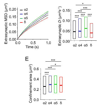
Our results indicate that GABAA receptors associated with phasic and tonic inhibition are both able to access inhibitory synaptic membranes by lateral diffusion, although subunits forming two major classes of extrasynaptic GABAA receptors (α5 and δ) are not retained at inhibitory synapses to the same extent as α2-and α4-GABAA receptors.

GABAA receptor subunits exhibit differential lateral mobility in the extrasynaptic membrane
Analysis of the membrane diffusion profiles for extrasynaptic GABAA receptors revealed that δ-GABAARs were the slowest to traverse the membrane, surprisingly α5-GABAARs were fastest, and α2-and α4-GABAARs were ranked intermediate.
The lower diffusion coefficients noted for extrasynaptic δ-GABAARs suggested that their mobility must be limited by diffusional constraints.
In extrasynaptic membrane areas, α2 receptors were most confined followed by δ, α4 and α5.
Lateral mobility and synaptic localization is determined by subunit composition and domains
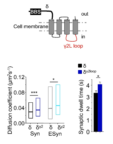
The second intracellular loop of γ2L subunits increased the trapping of GABAARs at inhibitory synapses.
The movement of δ subunits are controlled by diffusional constraints in the extrasynaptic membrane domain compared to other GABAAR subunits that have been studied in this investigation. The second intracellular loop of the δ subunit is important for reducing their lateral mobility and causing confinement to smaller areas of the extrasynaptic membrane.
For more details please visit the publication:
*corresponding authors
